+1 619-659-1180


Pre Purchase Exam
The actual pre-purchase examination is the most crucial expense the horse owner could make. The actual examination will help you choose if this particular horse is going to be right for you. Most of all, the actual pre-purchase will help the horse buyer from purchasing the wrong horse.
Obtain a written, signed history from the owner and/or agent selling the horse. The history should describe the horse, detail any previous medical and surgical problems, indicate the dates of any past radiographs (x-rays), list any medication given in the past 2 weeks, list dates of dewormings and vaccinations in the past year, and the pregnancy status of the horse, if a mare.
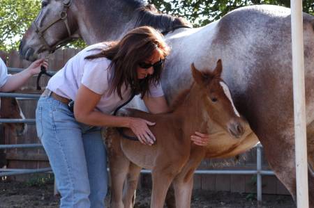
Here are examples of the pre-purchase exam
- We examine the horse’s general medical condition including temperature, eyes, coat, G.I. Auscult for sand, lungs, and heart.
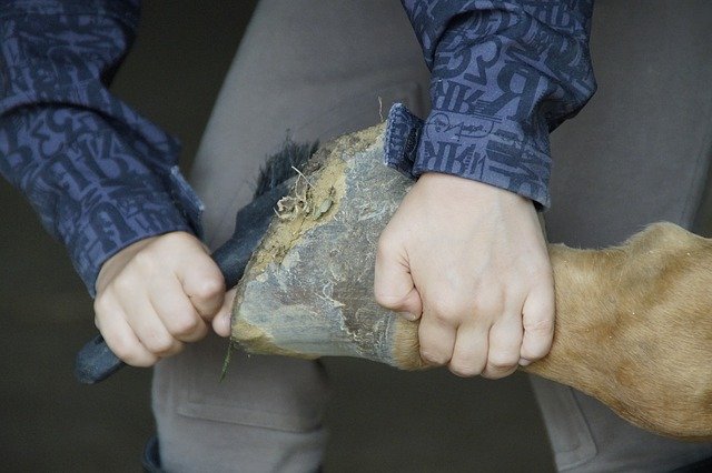
- The orthopedic portion of the examination, we palpate all tendons and joints flex and apply hoof testers to all four feet. We then walk, trot, and lunge the horse.
- The final evaluation, we would have the buyer of the horse ride the horse.
Additional Exams, with regard extra costs consist of
Radiographic (x-ray) examination associated with stifles, hocks, legs, fetlocks, as well as feet.
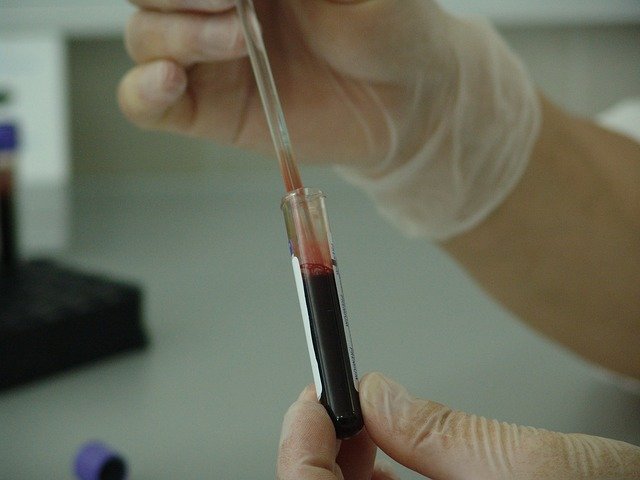
Blood Screens to evaluate for infection and anemia. A drug screen can detect any drugs in the horse’s system.
Reproductive exam including uterine culture or semen evaluation if the horse is going to be used for breeding.
Dr. Garfinkel will perform any of these tests or any other test you might be interested in having done. If you wish to have any of these test done contact us at 619.659.1180.
Confidentiality and Honesty
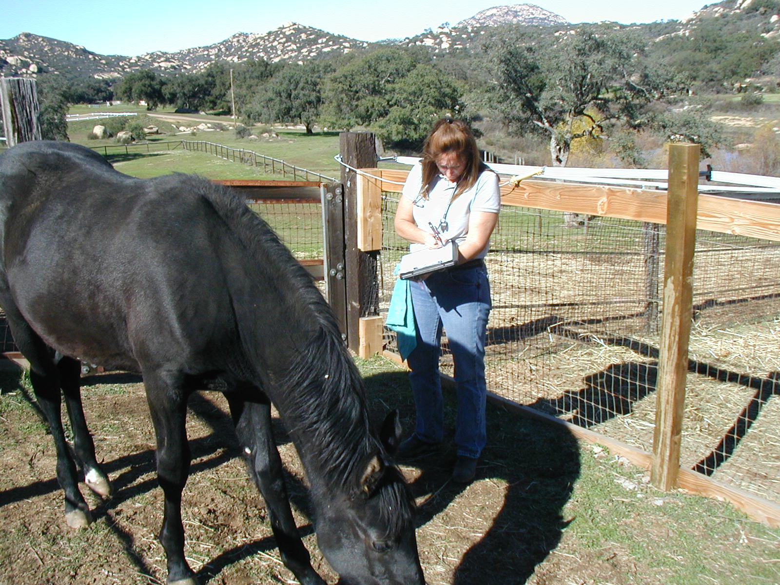
Generally when we do find a problem in the horse, the sellers were not aware of issue before the examination. The examination should be used to help you make an educated decision whether or not to buy the horse. Dr. Garfinkel respects the owner’s right to confidentiality, and you should to. She will not examine a horse for you (the buyer) if the seller is her client.
Dr. Garfinkel does not “pass” or “fail” any horse she examines. You the buyer will make the final decision to purchase or not to purchase the horse.
If you have any concerns or questions about Pre-Purchase Examinations contact us at 619.659.1180.
Lameness Exams
Lameness problems in horses are an ever-growing concern for horse owners. With a rising demand for top performance, the challenge is for veterinarians to keep up with improved diagnostics and treatment modalities. Furthermore, many work-ups today are related to improving performance, not just solving an obvious lameness issue, as subtle pain can hamper performance in any discipline.
It also should be mentioned that many of today’s top performance horses compete year-round without a break. This is tough for any athlete, and keeping horses comfortable and competing at a top level can be a huge challenge for any veterinarian.
This article will focus on how Dr. Garfinkel figure out what is causing problems when horses cannot tell us where it hurts.
The Initial Horse Exam
When working on lameness issues, almost always a veterinarian will "start at the bottom." Many lameness issues originate in the foot, so that is frequently the best place to start. Many times the trainer or rider will insist that the lameness is originating from the shoulder, when actually it is confined to the foot.
The first step in any lameness/poor performance examination for our practice is evaluating the horse's conformation and shoeing/trimming in relationship to the horse's discipline. From there, we perform a thorough exam of the foot, which includes using hoof testers to evaluate pain within the hoof.
We evaluate the foot by palpating the coronary band and feeling for any heat, pain, or swelling; palpating the heel bulb area; and squeezing the frog and sole of the foot with hoof testers. In addition, we palpate the digital arteries low on the heel to detect an increased pulse, if present. A positive finding to any of these can be a clue that inflammation is affecting the foot. For example, a localized soreness to an area of sole and an increased digital pulse can be an indication of a brewing hoof abscess, which can be explored further.
We also note any abnormalities in hoof balance or conformation at rest, then, when the horse is in motion, we watch such characteristics as how the horse's hooves land. Does one side of the hoof land first, or does the foot contact the ground toe first or heel first?
We palpate the limbs, including the tendons, ligaments, and joints, looking for areas of swelling, heat, or pain (the cardinal signs of inflammation). The horse's joints are then flexed, looking for any decrease in their range of motion or pain on flexion. This "physical" part of the exam is paramount in determining the source of a lameness, but it does not always provide a veterinarian with enough information to determine the source of a lameness.
Horse Movement
The next step in a lameness examination is to observe the horse walking and trotting in a straight line. Veterinarians are watching to see how the horse's limbs travel in motion. Do the limbs travel straight, or do they curve inward or outward before the feet land? Flexion tests are again performed, this time to observe whether or not the horse becomes lame and for how long after the flexion test. Flexion tests are performed to the joints of the lower limb of both the front and hind legs--the carpi (knees), the hocks, and the stifles--and often abduction tests are also performed (the limb is stretched away from the body). Wedge tests are also sometimes performed to the lower leg: a horse is stood with an angled plane (wedge) under one foot, then he is moved off at the walk or trot.
Flexion tests can be very useful in isolating a suspected source of lameness, but it should be noted that they are not the final answer. Some horses can flex test quite badly even without a significant abnormality within the joint, so these tests must be used as a piece of the puzzle and backed up by additional diagnostic information.
In many cases, veterinarians will want to watch the horse on the longe line and possibly under saddle. Some horses are not very lame in a straight line, but when asked to jog or trot on the longe line or to perform figure eights under saddle, the lameness will become much more evident. The type of surface can play a role in determining lameness. A hard surface, such as asphalt or concrete, will often expose a subtle lameness that is harder to notice on a softer surface.
The most important aspect when performing a lameness evaluation is: Which leg is the lame one?
When the horse is lame on a foreleg, watching the horse's head can be quite helpful to sort this out. In this case, when the lame leg is weight-bearing, the horse will usually raise his head upward in an effort to unload some weight off the lame leg. Then, as the sound leg becomes weight-bearing, the head will go down (many veterinarians are taught the expression "down on the sound leg" in veterinary school). Sometimes all that is evident is the horse's neck muscles become tense or that the lame leg spends less time on the ground and there is uneven loading of the fetlocks, or he might simply land harder on one leg, but these are all clues to which leg is causing the problem.
In the hind end, in moderate to severe lameness, horses will also raise and lower their heads to decrease the weight placed on the lame leg. So, with a right hind lameness at a trot, the horse will use his head as a lever to alleviate the load on the hind end, and he will appear to lower his head when the right hind leg is weight-bearing and raise his head when the left hind leg is weight- bearing. In essence, he'll mimic a right forelimb lameness.
In less severe cases, hind limb lameness is also determined from watching--among other things--the symmetry of movement of the horse's gluteal (rump) muscles. A horse with hind-limb lameness will not bear weight evenly, so gluteal rise and fall will not be even. A horse that is lame in one leg will have a shortened stride, so the gluteal rise will be shorter on one side than the other side when viewed from behind.
The horse will appear to have a "hip drop" from the shortened stride. Many veterinarians will also look for a "hip hike" in the lame leg during the early weight- bearing phase of the stride.
Watching the stride length of a hind-limb lameness will also sometimes point to the lame leg when the horse is viewed from the side.
Hind-limb lameness can be challenging, and every veterinarian has his or her own system for evaluating lameness. This is just a brief overview of some of the tools veterinarians use to help sort it out.
Making the Grade
During the course of a lameness examination, you might hear your veterinarian call a lameness by a certain grade. This helps standardize lameness severity and aims to make it easier when multiple veterinarians are involved and for recordkeeping. Some veterinarians just use the terms mild, moderate, or severe for grading lameness.
One Block at a Time
In some lame horses there is little or no evidence of inflammation--there is no obvious "sore spot" on a leg. Inthese cases local anesthesia (called nerve blocks) can be performed to definitively localize the area of pain (lameness) so that further diagnostic tests can be performed.
In horses, nerves travel down the inside and outside (medial and lateral) of the legs, like electrical wires, branching off to more and more specific areas of the leg. Local anesthetic can be deposited around the nerves at specific levels, thereby anesthetizing the area. After waiting anywhere from five to 15 minutes (it's the veterinarian's preference), the horse is reevaluated. If the horse becomes sound or the lameness is significantly reduced (by at least 50%), then something in that area is suspected of causing the lameness and imaging studies (radiographs, ultrasound, CT scan, or MRI--more on these below) can be performed. In some cases initial imaging studies will not determine the source of a lameness, so, if suspected, an individual joint or tendon sheath can be "blocked" for an even more precise determination of where it hurts.
What's Inside Equine Lameness
Once the determination is made as to what area is causing the lameness, the veterinarian can utilize imaging studies to determine the true cause of the lameness and institute a treatment plan. The most common imaging tools for lameness evaluation are radiography and ultrasound. Radiography is for evaluating bone structures and is quite useful in detecting issues such as arthritis and/or fractures. Ultrasound, on the other hand, is better for evaluating soft tissue structures by using sound waves to create a picture based on density. All soft tissues have unique textures and densities that appear as specific patterns on the ultrasound image with disruption of the normal patterns being indicative of injury.
Computed tomography (CT) or MRI are two of the latest imaging tools to help veterinarians determine causes of more troublesome lameness spots, such as caudal (toward the rear) heel pain or other lamenesses confined to the foot that are not well-defined on radiographs or ultrasound.
In some cases, nerve blocks are not performed due to risk of injury or because the patient is uncooperative. In these cases, nuclear scintigraphy (bone scan) on the sedated horse can be performed at a hospital to help determine the cause of a lameness.
Take-Home Message
Dr. Garfinkel has more tools to help diagnose and treat specific lameness problems. If your horse is experiencing a problem, contact Dr. Garfinkel at 619.659.1180 sooner rather than later for the best chance of diagnosing and correcting the problem.