+1 619-659-1180


HORSE NAVICULAR DISEASE
Navicular disease, also called navicular syndrome or podotrochleosis, describes a number of problems affecting the distal sesamoid or navicular bone in the foot. Navicular disease is a responsible for approximately one-third of all chronic lameness in horses. The condition comprises complex of problems. Some of these problems are completely reversible, some can be improved with appropriate long-term management, and some are irreversible.
Conditions related to navicular disease include coffin joint arthritis, deep flexor bursitis and/ortendonitis, inflammation of the ligaments of the navicular bone, and problems relating to thenavicular bone itself (bone spurs, fractures, cysts), its blood supply, and hoof changes.
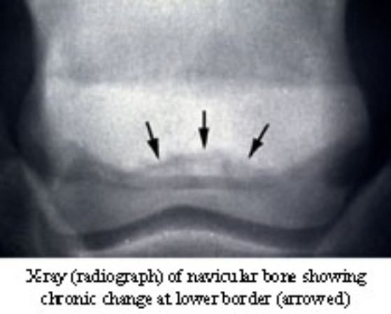
The boat-shaped ("navy") navicular bone is located behind the juncture of the pastern bone (P2) and the coffin bone (PI), at about the level of the coronet; it is thus part of the coffin joint. The navicular bone acts as a fulcrum over which the deep flexor tendon moves. The bone is embeddedin and surrounded by ligaments, and is almost impossible to expose during surgery. It is difficult to place a needle into the coffin joint, and it is difficult to obtain high-quality radiographs (x-rays) of the area.
Numerous factors affect the health of the navicular bone and related coffin joint:
- Abnormal conformation: A small foot attached to an upright pastern; a long-toed, low-heeled foot; mismatched feet that become more of a problem as the horse ages.
- Foot carriage: The way the foot lands puts unequal pressure on the navicular area; loading one side of the foot more than the other; landing more toward the toe or heel.
- Shoeing: This may cause more weight to be carried on one leg than on another; shoe size, typeand position and the shoeing schedule influence navicular bone health; a shoe must bl~large enough to provide adequate support, especially in the heel area; irregular shoeing createsdifferent foot angles to which the coffin joint must adjust with exercise. This stresses the joint.
- Hoof wall quality, compliance and conformation: A hoof wall that is too hard, too soft, too thin, crumbly or cracked.
- Exercise: Lack of daily regular exercise; irregular exercise; work beyond the horse's state of conditioning; exercise on hard, unforgiving surfaces; speed at which the horse travels; weight of the rider, tack and the horse's body itself.
- Diagnosis and management: Navicular disease can be very frustrating to diagnose and treat, both for the veterinarian and the owner. Your patience as an owner is very important for successful management of the problem. We may need to try various treatments, such as different shoeing, medica- tion and exercise programs to observe your horse's response to diagnose navicular disease. Diagnostic methods include a thorough orthopedic examination (probably more than one), local anesthesia (nerve blocks), radiographs (x-rays) and other diagnostic tests.